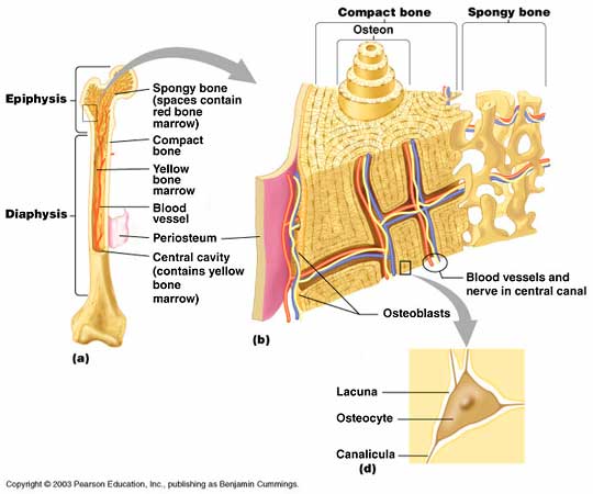
4. List, describe, and illustrate the major anatomical areas of a long bone.
Epiphyses
- There are two epiphyses of a long bone: the proximal epiphysis and the distal epiphysis
- The epiphyses are the ends of the long bone
- They consist of a thin layer of compact bone that surrounds an area of spongy bone
- Articular cartilage covers the epiphyses, rather than the periosteum that covers the diaphysis
- The articular cartilage is benefitical in joint movement because it decreases the friction between bones/joints
- Within the spongy bone inside the epiphyses lies the epiphyseal line, a thin line of bony tissue that used to be the epiphyseal plate (an area for growth in young bones, where hyaline cartilage used to be)
Diaphysis
- Shaft, makes up most of the bone's length
- Composed of compact bone
- Covered by the periosteum
- The periosteum is attached to the bone by perferating (or Sharpey's) fibers
- Within the diaphysis is the medullary cavity, which stores red and yellow bone marrow
- Red bone marrow (red blood cells) is typically found in the medullary cavity of infants
- Yellow bone marrow (fat cells) is more present in adult bones
More information about long bones: http://visual.merriam-webster.com/human-being/anatomy/skeleton/structure-long-bone.php
5. Explain the chemistry involved in making the bone both hard and flexible.
- There is a collagen matrix (a fibrous protein component of bone) that makes up about 35% of bone
- The matrix stores a lot of protein and calcium
- This gives the bone flexibility
- The matrix also collects calcium and phosphorous salts which give the bone strength
For more information about the strength and flexibility of bones (and a great recipe to promote strong, healthy bones) click here!
